OPHTHALMOLOGY
Tonometer safely checks pet’s eyes
 What is glaucoma?
Glaucoma is a disease of the eye in which the pressure within the eye, called the intraocular pressure (IOP) is increased. Intraocular pressure is measured using an instrument called a tonometer.
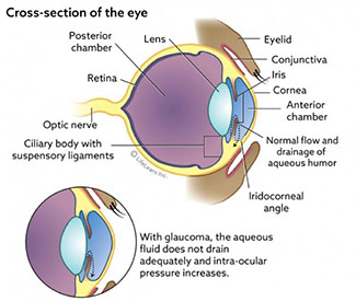
What is intraocular pressure and how is it maintained?
In addition to producing this fluid (aqueous humor), the ciliary body contains the suspensory ligaments which hold the lens in place. Muscles in the ciliary body pull on the suspensory ligaments, controlling the shape and focusing ability of the lens.
Aqueous humor contains nutrients and oxygen that are used by the structures within the eye. The excess fluid is constantly drained from the eye between the cornea and the iris. This area is called the iridocorneal angle, or the filtration/drainage angle.
The intra-ocular pressure remains constant as long as the production and absorption or drainage of aqueous is equal.
What causes glaucoma?
Glaucoma is caused by inadequate drainage of aqueous fluid; it is not caused by overproduction of fluid. Glaucoma is further classified as primary or secondary glaucoma.
Primary glaucoma results in increased intra-ocular pressure in a healthy eye. Some breeds are more prone than others (see below). It occurs due to inherited anatomical abnormalities in the drainage angle.
Secondary glaucoma results in increased intra-ocular pressure due to disease or injury to the eye.
This is the most common cause of glaucoma in dogs and cats. Causes include:
- Uveitis (inflammation of the interior of the eye) or severe intra-ocular infections, resulting in debris and scar tissue blocking the drainage angle.
- Anterior dislocation of lens. The lens falls forward and physically blocks the drainage angle or pupil so that fluid is trapped behind the dislocated lens.
- Tumors can cause physical blockage of the iridocorneal angle.
- Intra-ocular bleeding. If there is bleeding in the eye, a blood clot can prevent drainage of the aqueous humor.
- Damage to the lens. Lens proteins leaking into the eye as a result of a ruptured lens can cause an inflammatory reaction resulting in swelling and blockage of the drainage angle.
Are certain breeds more likely to get glaucoma?
Yes, the following breeds are associated with glaucoma:
Akita Dalmatian Norwegian Elkhound Alaskan Malamute English Cocker Spaniel Poodle American Cocker Spaniel English Springer Spaniel Samoyed Basset Hound Flat-Coated Retriever Shar Pei Beagle Giant Schnauzer Shih Tzu Boston Terrier Great Dane Siberian Husky Bouvier des Flandres Greyhound Smooth-Haired Fox Terrier Bull Mastiff Italian Greyhound Welsh Springer Spaniel Chow Chow Miniature Pinscher Wirehaired Fox Terrier Cocker Spaniel Miniature Schnauzer
Cats can get glaucoma too!
Feline aqueous humor misdirection syndrome (FAHMS or “malignant glaucoma”). This unique disorder has been described in older cats (average age 11.7 years), with a greater incidence in females. Some ophthalmologists note that Persians are overrepresented. Characteristic signs include anisocoria (two different sized pupils), with relative mydriasis (dilated pupil) of the affected eye, sluggish pupillary light reflexes, and a uniformly shallow anterior chamber. The disease is linked to aqueous humor misdirection into the vitreous. The expanding vitreous volume displaces the lens and iris forward. IOP increases as the iridocorneal angle narrows and the ciliary cleft collapses.
Most feline glaucomas, however, are secondary to chronic intraocular inflammation and can be especially difficult to treat given the dearth of effective anti-glaucoma medications in this species. Typically, uveitis leads to a secondary glaucoma in cats.
Why is an increase in intraocular pressure a problem?
High intraocular pressure causes damage or degenerative changes to occur in the retina and the optic nerve. The retina is the innermost lining layer of the eyeball, located on the back surface. It contains the light-sensitive rods and cones and other cells that convert images into nerve signals. The optic nerve is the nerve that runs from the back of the eye to the brain, and it carries the signals from the retina to the brain, thus producing vision.

What are the signs of glaucoma and how is it diagnosed?
The most common signs noted by owners are:
- Eye pain. Your dog may partially close and rub the eye. He may turn away as you touch him or pet the side of his head.
- Watery discharge from the eye.
- Lethargy, loss of appetite, or even unresponsiveness.
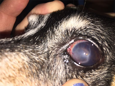
- Obvious physical swelling and bulging of the eyeball. The white of the eye (sclera) looks red and engorged.
- The cornea or clear part of the eye may become cloudy or bluish in color.
- Blindness can occur very quickly unless the increased IOP is reduced.
All of these signs can occur very suddenly with acute glaucoma. In chronic glaucoma they develop more slowly. They may have been present for some time before your pet shows any signs of discomfort or clinical signs.
“Acute glaucoma is an emergency.”
Diagnosis of glaucoma depends upon accurate IOP measurement and internal eye examination using special instruments. Acute glaucoma is an emergency. Sometimes immediate referral to a veterinary ophthalmologist is necessary.
How is it diagnosed?
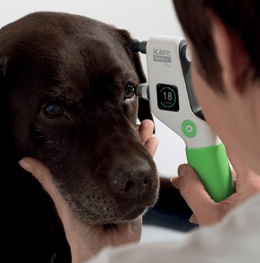
Tonometry is the procedure performed to determine intraocular pressure (IOP), the fluid pressure inside the eye. The measurement of IOP is used in the diagnosis of glaucoma and other ocular diseases.
At TOVWC we use a state-of-the-art tool called the TONOVET Plus Tonometer to safely and effectively check the IOP of your pets’ eyes. Icare® TONOVET Plus is the new generation tonometer for quick and easy veterinary IOP measuring. The gentle measurement does not require any sedation or pain and typically takes less than five minutes to measure.

What is the treatment for glaucoma?
It is important to reduce the IOP as quickly as possible to reduce the risk of irreversible damage and blindness. It is also important to treat any underlying disease that may be responsible for the glaucoma.
“It is important to reduce the IOP as quickly as possible to reduce the risk of irreversible damage and blindness.”
Analgesics are usually prescribed to control the pain and discomfort associated with the condition. Medications that decrease fluid production and promote drainage are often prescribed to treat the increased pressure. Long-term medical therapy may involve drugs such as carbonic anhydrase inhibitors (e.g., dorzolamide 2%, brand names Trusopt® and Cosopt®) or beta-adrenergic blocking agents (e.g., 0.5% timolol, brand names Timoptic® and Betimol®).
Medical treatment often must be combined with surgery in severe or advanced cases. Veterinary ophthalmologists use various surgical techniques to reduce intra-ocular pressure. In some cases that do not respond to medical treatment or if blindness has developed, removal of the eye may be recommended to relieve the pain and discomfort.
Is follow-up treatment necessary?
Once glaucoma is diagnosed and medication is started, follow-up monitoring will be necessary. Initially, your veterinarian will recommend frequent follow-up examinations to ensure that your dog is responding adequately to the treatment or to make adjustments to the medications.
What is the prognosis?
The prognosis depends to a degree upon the underlying cause of the glaucoma. In the long-term, constant medical treatment will be required to keep the disease under control.
If you own a dog on the predisposed list of breeds you should have glaucoma testing done at the time of the annual physical examination.
If you see any of the signs mentioned above in your dog or cat, please call the office immediately as early detection of glaucoma may make the difference between losing and saving their vision.
